
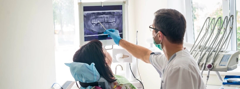
Digital Dental X-Ray Clinic Near You In Bikaner
If you have been searching for an OPG dental x ray near you or a reliable dental x-ray in Bikaner, you are in the right place. At Dencity, digital OPG imaging gives your dentist a single, full-picture view of your teeth, jaws, and surrounding bone, making diagnosis faster, clearer, and more precise than ever before.
What Exactly Is A Digital OPG X-Ray?
Simply said, OPG stands for Orthopantomogram, a panoramic dental X-ray that captures the entire oral and maxillofacial region in one continuous, two-dimensional image. Unlike conventional radiographs that focus on individual teeth, an OPG covers both upper and lower jaws (maxilla and mandible), all erupted and unerupted teeth, the temporomandibular joints (TMJ), and the surrounding alveolar bone simultaneously.
The technology behind it is straightforward. A rotating arm moves around the head while a digital sensor sweeps the opposite side, recording image data in real time. The result is a single composite dental radiograph that would otherwise require several separate exposures to replicate. If you are someone who’s looking for a dental X Ray clinic near you with full-mouth diagnostic capability, this is the standard of care.
- ≤15-Second Scan Time: The scanner takes rapid images, from patient positioning to final capture in just 15 seconds or less.
- ~80% Radiation Reduction: Significantly safer than older, traditional film-based systems.
- Single-Shot Exposure: Provides comprehensive, full-mouth coverage in just one take.
- Instant Availability: Images are ready for immediate review within seconds.
Step-By-Step Look At How An OPG Scan Works?
The process is non-invasive, takes under two minutes from start to finish, and requires no injections or special preparation. Here is what happens during a standard OPG scan:
- Positioning And Chin Rest: The patient is comfortably positioned with their chin resting on the support.
- Bite Peg Alignment: A tiny bite peg makes sure the jaw alignment is proper and prevents overlapping.
- Arm Rotates And Scans: The arm moves smoothly around the head, capturing everything it needs.
- Digital Image Compiles: Finally a top notch software instantly compiles all that info into one clear digital image.
- Doctor Reviews And Diagnoses: The practitioner evaluates the complete scan for immediate diagnosis.
Jewelry or any metallic accessories near the head must be removed beforehand, as they create artifacts on the image. Pregnant individuals should inform the clinic prior to the procedure. Other than these precautions, there is nothing further required from the person being scanned.
What Gets Captured In An Intraoral X-Ray (Inside the Mouth)?
As an Intraoral X-Ray clinic in Bikaner, Dencity offers both major intraoral imaging modalities, each suited to different diagnostic needs. In intraoral radiography, the sensor is placed directly inside the mouth to capture close-range detail of specific teeth or a localized region.
- Bitewing X-Ray: This shows both the upper and lower posterior teeth together, and is especially good for spotting any decay between adjacent teeth, which just can't be spotted with the naked eye. It also checks the height of the alveolar bone, making it super helpful for diagnosing periodontal issues early.
- Periapical (Periradicular) X-Ray: It presents the entire tooth length, from crown to root tip and even the surrounding bone. This is used a lot in case of checking root canals, finding periapical abscesses, and assessing bone quality around implants.
- Occlusal X-Ray: It provides a wider view of the whole dental arch, either it can be upper or lower, in one shot. This is helpful for finding extra teeth, spotting jaw fractures, and keeping track of tooth development in kids.
Extraoral X-Ray: Full-Picture Views For Complex Diagnoses
Dencity functions as a dedicated Extraoral X-Ray clinic in Bikaner, the imaging here extends well beyond just individual teeth. Extraoral radiography places the sensor outside the mouth and is used when the clinical question involves the entire jaw, facial skeleton, or TMJ region.
- OPG/Panoramic Radiograph: This is super common and also handy. It shows all the teeth, both jaw arches, TMJ, and the maxillary sinuses at once. It is known to be really helpful for orthodontics, checking wisdom teeth, and planning full arch implants too.
- Lateral Cephalometric Projection: The lateral cephalometric projection shows the skull in profile. It's mainly used in orthodontics and jaw surgery to see how teeth, jaws, and the cranial base relate. This is really important for planning treatments in complex malocclusion cases.
- Multi-Slice CT/CBCT: Multi-slice CT or CBCT provides detailed 3D cross sections of maxillofacial area. When normal 2D views aren't enough for complex implant placements or TMJ problems that need volumetric data, this method is used.
When Does Your Dentist Recommend An OPG Scan?
Not every dental concern requires a panoramic view but several conditions genuinely benefit from it. If you are looking for the nearest dental x ray centre because your dentist has recommended imaging, the table below covers the most common clinical scenarios where an OPG is the first choice.
| Clinical Scenario | Why Is OPG Recommended? |
|---|---|
| Wisdom tooth evaluation | Assesses angulation, depth, and proximity to the inferior alveolar nerve before extraction |
| Orthodontic treatment planning | Maps tooth development, eruption sequence, and jaw proportions for bracket or aligner prescriptions |
| Dental implant workup | Evaluates available bone height and width; identifies sinus floor proximity |
| TMJ pain or clicking | Visualizes joint morphology and detects degenerative changes in condylar head |
| Suspected jaw cyst or tumor | Reveals radiolucent or radio-opaque lesions within the jaw that are not visible clinically |
| Children's tooth development review | Tracks mixed dentition, detects congenitally missing teeth, and anticipates eruption problems |
| Post-treatment monitoring | Evaluates healing after bone grafts, surgical extractions, or endodontic treatment |
Comparison Of Digital And Conventional X-Ray
If you are weighing your options and want the best dental x-ray in Bikaner, understanding what makes digital technology clinically superior to traditional film-based systems is important. The differences go beyond convenience.
| Parameter | Digital OPG | Conventional Film X-Ray |
|---|---|---|
| Radiation dose | Up to 80% lower | Significantly higher |
| Image availability | Instant, seconds after scan | Requires darkroom development |
| Image enhancement | Contrast, zoom, brightness adjustable | Fixed, no post-processing |
| Storage and sharing | Digital: cloud, email, referral-ready | Physical film; deteriorates over time |
| Early decay detection | High sensitivity even at small sizes | Limited by film grain and contrast |
| Environmental impact | No chemical processing required | Uses chemical developers and fixers |
Conditions That A Digital OPG Helps Detect And Diagnose
A digital OPG is not simply a screening tool, it is a diagnostic workhorse. At our dental x-ray clinic in Bikaner, the imaging supports detection and assessment of a wide range of oral and maxillofacial conditions, many of which present no obvious symptoms in early stages.
The following are conditions routinely identified through panoramic dental X-ray evaluation:
- Impacted wisdom teeth
- Jaw bone loss (periodontitis)
- Dentigerous and radicular cysts
- Supernumerary (extra) teeth
- Jaw fractures
- Ameloblastoma and jaw tumors
- Maxillary sinus pathology
- TMJ degeneration
- Interproximal dental caries
- Root resorption
- Periapical abscesses
- Developmental anomalies
Because the image captures the full arch in one frame, the radiologist can also pick up incidental findings, such as calcified carotid atheromas or salivary gland calculi that would otherwise go unnoticed in a targeted intraoral view.
What The Numbers Mean In Radiation Exposure In OPG Imaging?
Radiation concern is one of the most common questions people raise before agreeing to any dental imaging. It is a fair question, and one worth answering with accuracy rather than reassurance.
A digital OPG does the work with an effective dose of approximately 14–24 microsieverts (µSv), roughly equal to the natural background radiation that a person receives over two to four days of daily living.
For example a chest X-ray exposes you to around 100 µSv. Digital systems lower this because of the optimized sensor sensitivity and collimated beam geometry. This lets dental imaging use really low radiation levels, making it more than just a marketing line.
Clinical Note: The ALARA principle (As Low As Reasonably Achievable) governs all radiographic decisions at Dencity. An X-ray is recommended only when the diagnostic benefit outweighs the negligible exposure involved not as a routine procedure without clinical indication.
Why Dencity Is Preferred Place For The Best Dental X-Ray Clinic In Bikaner?
Picking a dental imaging service isn't just about convenience. The image accuracy, gear quality, and how well it fits with your overall care plan really count too. At Dencity, they integrate your imaging right into your treatment plan, no outsourcing or looking at things in isolation.
Dr. Ajay Singh Rajpurohit (MDS, Orthodontics and Dentofacial Orthopaedics) and Dr. Divya Rajpurohit (MDS, Pedodontics and Preventive Dentistry) both trained and bring specialist level reading of dental radiographs to every case. Whether the imaging is for an orthodontic workup, a suspected cyst, or a pre-implant jaw bone assessment, the findings are reviewed in the context of a full clinical examination, not handed back as a standalone film.
Ready To Book Your OPG Dental X Ray At Dencity?
As a full-service dental x-ray clinic in Bikaner, Dencity operates modern digital OPG equipment that delivers high-resolution images with minimal exposure. For those specifically seeking the best dental x-ray in Bikaner, the combination of specialist interpretation and calibrated imaging technology makes a meaningful clinical difference.
Walk in or call ahead, our team is the best for dental X Ray in Bikaner, and will guide you to the right imaging based on what your dentist has recommended. Same-day imaging appointments are available.
Book an Appointment right away...